In the fight against oral squamous cell carcinoma (OSCC), a prevalent and malignant cancer with a survival rate of only 50% – 60%, early and accurate diagnosis is critical for improving patient outcomes. At Tianjin University of Traditional Chinese Medicine, Zhang et. al. employ microscopic hyperspectral imaging and machine learning (ML) to develop a more effective method of tissue examination during pathological staging. Currently, pathological staging is limited by the examiner’s ability to visually recognize cancer indicators in cell and tissue structure, with only a microscope, their eyesight, and their experience. By implementing hyperspectral imaging and machine learning, Zhang et al. hope to further regulate this process and minimize any errors in diagnoses by using spectral identification to limit human error.
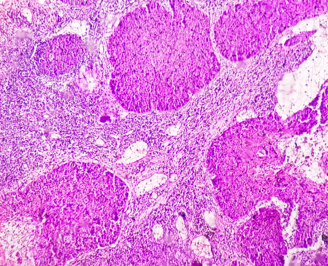
As cancer develops and progresses, the cellular structure and the chemical makeup are altered. Though imperceivable to the human eye, the spectral changes caused by the altered chemistry are visible when imaged by a hyperspectral camera. Hyperspectral imaging looks at the spectra of a very narrow region of light in high detail, with chemical identification of a sample possible through analysis of the width, amplitude, and wavelength of absorption peaks. These images are three-dimensional data cubes, with two spatial dimensions and one spectral dimension, offering a spectrum at each point in an image as seen in Figure 1. Hyperspectral imaging is an extraordinarily powerful tool that, when combined with microscopy and machine learning, can be used to analyze the health and integrity of cells and offer highly accurate staging prognoses.

Zhang et. al. mounted a SOC710-E, a hyperspectral camera with a range of 400 – 1000 nanometers, onto their 40x biological microscope (E100, Nikon) to take these images. The SOC 710E offers internal line scanning and a C-mount, which makes it highly suitable for microscopy applications. Though hyperspectral microscopy enables spectral analysis and differentiation of cells, the differences in the spectra can still be subtle.
As such, Zhang et. al. utilize a variety of preprocessing methods, data dimensionality reduction strategies, and classification models to determine the most efficacious analysis procedure. The effectiveness of the different models was determined by the accuracy in diagnosing cells as either in the well-differentiated stage, which is an earlier stage with minimal differences from normal cells, the moderately-differentiated stage, which has a larger difference from normal cells, or the poorly-differentiated stage, with a large difference from normal cells. These diagnoses were made based on the spectra of each cell sample. Figure 2 displays the averaged spectra of each pathological stage.

Ultimately, two of their models resulted in 100% accuracy, proving that combining microscopic hyperspectral imaging with machine learning is a successful method of pathological staging. While further work is needed to streamline the data reduction and analysis process, this research lays the groundwork for more accurate and efficient diagnoses. Zhang et. al. hope that this will ultimately increase the survival rate of those afflicted with OSCC.
Full Publication
Zhang, Yuanhao and Liu, Zhaowei and Wu, Chenlu and Liu, Ming and Li, Gang and Han, Xiangli and Suo, Tongchuan and Zhao, Jing, Study on Determining the Pathologic Staging of the Oral Squamous Cell Carcinoma Based on Microscopic Hyperspectral Imaging. Available at: http://dx.doi.org/10.2139/ssrn.5120876
Ready to Explore Hyperspectral Imaging for Your Application?
Curious if hyperspectral imaging could work for your project? Drop us a line—we’d love to hear what you’re working on.