
Tattoos have become an increasingly popular form of body art, with an estimated 32% of Americans having at least one. However, when it comes to removal, there is still no simple or guaranteed solution. Laser removal remains the most common method, but fully eliminating pigment can be difficult. Recent research from the Medical University of Silesia highlights how hyperspectral imaging technology can be used to analyze the optical properties of tattoo inks—paving the way for more effective, targeted treatment strategies.
The Challenge of Current Tattoo Removal Methods
Current laser tattoo removal is based on the principle of selective photothermolysis, where laser energy is absorbed by tattoo pigments while minimizing damage to the surrounding skin. Achieving this selective absorption is critical to ensuring both the safety and effectiveness of the procedure. However, in addition to tattoo dyes, laser energy can also be absorbed by endogenous skin chromophores—primarily water, hemoglobin, and melanin—potentially leading to unintended thermal damage. For optimal results, the laser wavelength must be carefully chosen to maximize absorption by the target pigment while minimizing interaction with surrounding tissue. Because different ink colors have distinct absorption peaks, effective treatment often requires the use of lasers operating at multiple wavelengths.
Experimental Design
The aim of the study conducted by the Medical University of Silesia was to investigate the spectral properties of dyes in order to maximize laser radiation absorption by pigments. The research team analyzed seven commercially available tattoo dyes sourced from established manufacturers. Sample preparation involved applying two drops of each dye to glass slides and spreading them uniformly across the surface. After drying, researchers subjected each sample to hyperspectral analysis using the SOC-710 system with incandescent illumination. The SOC710-VP operates in the 400-1000 nm spectral range, with a spectral resolution of 4.69 nm and a spatial resolution of 696 pixels per line.

Key Findings
- Maximum Reflectance Wavelengths: All tested dyes exhibited maximum reflectance between 634-732 nm, suggesting that lasers operating within this range would be least effective for removal. The red dye showed maximum reflectance at 634 nm, while the brown dye peaked at 732 nm.
- Reflectance Magnitude Variations: Perhaps most striking were the dramatic differences in reflectance intensity between dyes. The white pigment demonstrated maximum reflectance of 3,222 relative units, while the black dye measured only 85 relative units. This finding suggests that black tattoos should be significantly easier to remove than those with white or yellow pigments.
- Similar Colors, Different Spectral Properties: The research revealed that visually similar dyes can possess dramatically different spectral characteristics. Red dye showed reflectance of 751 relative units, while dark pink dye measured 1,304 relative units—nearly double the reflectance despite appearing closely related in color.
Reflectance Spectra of Tattoo Dyes
Along with wavelength selection, reflectance is a key factor in determining the effectiveness of laser tattoo removal treatments.
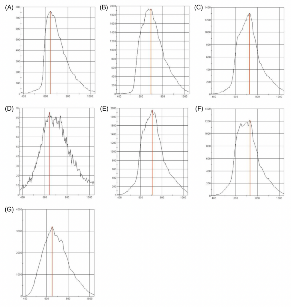
Reflectance spectra of tested tattoo dyes: red (A), yellow (B), dark pink (C), black (D), light pink (E), brown (F), and white (G).
Clinical Implications and Treatment Optimization
The research findings suggest several strategies for improving laser tattoo removal outcomes. Rather than selecting laser wavelengths based on visual color assessment, practitioners could optimize treatment by avoiding each dye’s maximum reflectance wavelength. For the tested samples, this means avoiding the 634-732 nm range and selecting wavelengths where absorption exceeds reflectance.
If tattoo ink manufacturers verify the spectral properties of dyes and include this information on the packaging, it will enable the optimization of laser radiation parameters. While these findings provide valuable insights, it is essential to note that the conducted analyses were performed in vitro on glass slides rather than in skin tissue.
Future research might explore non-invasive hyperspectral imaging of actual tattoos to validate these laboratory findings.
Full Publication
Stolecka-Warzecha A, Chmielewski Ł, Wilczyński S, Koprowski R. In vitro hyperspectral analysis of tattoo dyes.
Skin Res Technol. 2023; 29:e13268. https://doi.org/10.1111/srt.13268
Ready to Explore Hyperspectral Imaging for Your Application?
Curious if hyperspectral imaging could work for your project? Drop us a line—we’d love to hear what you’re working on.
Tattoos have become an increasingly popular form of body art, with an estimated 32% of Americans having at least one. However, when it comes to removal, there is still no simple or guaranteed solution.
Laser removal remains the most common method, but fully eliminating pigment can be difficult. Recent research from the Medical University of Silesia highlights how hyperspectral imaging technology can be used to analyze the optical properties of tattoo inks—paving the way for more effective, targeted treatment strategies.
The Challenge of Current Tattoo Removal Methods
Current laser tattoo removal is based on the principle of selective photothermolysis, where laser energy is absorbed by tattoo pigments while minimizing damage to the surrounding skin. Achieving this selective absorption is critical to ensuring both the safety and effectiveness of the procedure.
However, in addition to tattoo dyes, laser energy can also be absorbed by endogenous skin chromophores—primarily water, hemoglobin, and melanin—potentially leading to unintended thermal damage. For optimal results, the laser wavelength must be carefully chosen to maximize absorption by the target pigment while minimizing interaction with surrounding tissue. Because different ink colors have distinct absorption peaks, effective treatment often requires the use of lasers operating at multiple wavelengths.
Experimental Design
The aim of the study conducted by the Medical University of Silesia was to investigate the spectral properties of dyes in order to maximize laser radiation absorption by pigments. The research team analyzed seven commercially available tattoo dyes sourced from established manufacturers.
Sample preparation involved applying two drops of each dye to glass slides and spreading them uniformly across the surface. After drying, researchers subjected each sample to hyperspectral analysis using the SOC-710 system with incandescent illumination. The SOC710-VP operates in the 400-1000 nm spectral range, with a spectral resolution of 4.69 nm and a spatial resolution of 696 pixels per line.
Key Findings
- Maximum Reflectance Wavelengths: All tested dyes exhibited maximum reflectance between 634-732 nm, suggesting that lasers operating within this range would be least effective for removal. The red dye showed maximum reflectance at 634 nm, while the brown dye peaked at 732 nm.
- Reflectance Magnitude Variations: Perhaps most striking were the dramatic differences in reflectance intensity between dyes. The white pigment demonstrated maximum reflectance of 3,222 relative units, while the black dye measured only 85 relative units. This finding suggests that black tattoos should be significantly easier to remove than those with white or yellow pigments.
- Similar Colors, Different Spectral Properties: The research revealed that visually similar dyes can possess dramatically different spectral characteristics. Red dye showed reflectance of 751 relative units, while dark pink dye measured 1,304 relative units—nearly double the reflectance despite appearing closely related in color.
Reflectance Spectra of Tattoo Dyes
Along with wavelength selection, reflectance is a key factor in determining the effectiveness of laser tattoo removal treatments.
Reflectance spectra of tested tattoo dyes: red (A), yellow (B), dark pink (C), black (D), light pink (E), brown (F), and white (G).
Clinical Implications and Treatment Optimization
The research findings suggest several strategies for improving laser tattoo removal outcomes. Rather than selecting laser wavelengths based on visual color assessment, practitioners could optimize treatment by avoiding each dye’s maximum reflectance wavelength. For the tested samples, this means avoiding the 634-732 nm range and selecting wavelengths where absorption exceeds reflectance.
If tattoo ink manufacturers were to verify and disclose the spectral properties of dyes and include this information on the packaging, it would enable the optimization of laser radiation parameters. While these findings provide valuable insights, it is essential to note that the conducted analyses were performed in vitro on glass slides rather than in skin tissue.
Future research might explore non-invasive hyperspectral imaging of actual tattoos to validate these laboratory findings.
Access Full Publication
Ready to Explore Hyperspectral Imaging for Your Application?
Curious if hyperspectral imaging could work for your project? Drop us a line—we’d love to hear what you’re working on.