The Challenge of Monitoring Coral Reefs
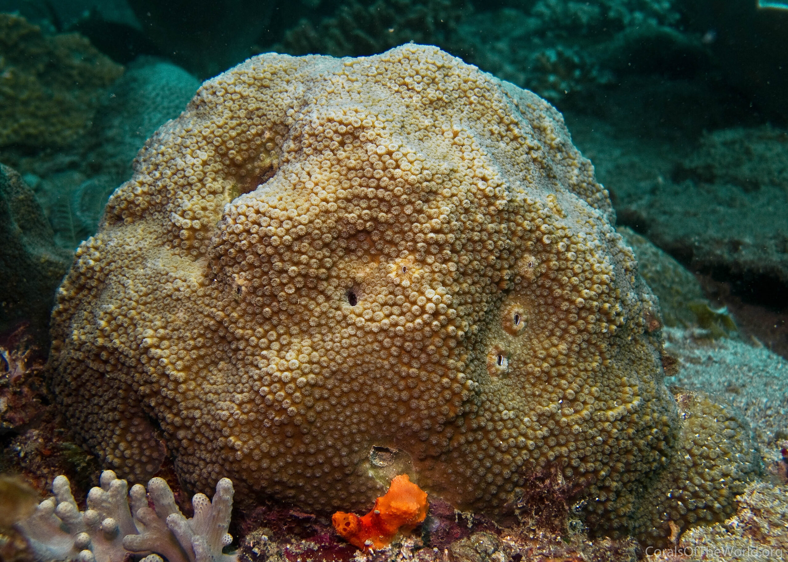
Coral reefs are among the most vibrant and ecologically significant ecosystems on Earth. However, they are also under threat due to changing ocean temperatures, pollution, and ocean acidification. Understanding how corals respond to environmental stressors is crucial for conservation efforts, but traditional methods of monitoring reef health are often time-consuming and invasive.
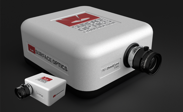
A collaborative study between the University of Connecticut, Pennsylvania State University, University of Delaware, and the University of Georgia has leveraged hyperspectral imaging to analyze coral reflectance and assess their health non-invasively. One of the key tools in this study was the 710-VP hyperspectral imager, manufactured by Surface Optics, which provided detailed spectral data that revealed subtle changes in coral pigmentation and bleaching.
What is Hyperspectral Imaging?
Hyperspectral imaging (HSI) is a cutting-edge technique that captures light across a wide range of wavelengths, far beyond what the human eye can perceive. Instead of just recording red, green, and blue light like conventional cameras, HSI systems generate a full spectrum of data for each pixel in an image. This allows researchers to detect minute differences in color and composition that indicate physiological changes in corals.
To learn more about hyperspectral Imaging, explore our spectral imaging resources section.
How the Study Was Conducted
To investigate coral reflectance, researchers used the 710-VP hyperspectral imager, which collects spectral data from 380 to 1040 nm at 5 nm increments. This system was mounted on a tripod and positioned above coral fragments submerged in a controlled tank environment. The corals were subjected to different temperature treatments to simulate heat stress and mimic real-world climate change effects.
Each coral sample was imaged under natural sunlight, and the hyperspectral data cubes were processed to generate detailed reflectance maps. These maps revealed how light interacts with coral tissue, providing insights into pigment concentrations and the presence of symbiotic algae known as zooxanthellae.
Key Findings: The Spectral Signature of Coral Bleaching
One of the most striking observations was the spectral response of corals under heat stress. Healthy corals typically exhibit a high absorption of light due to the presence of symbiotic algae, which play a vital role in photosynthesis. However, when corals experience stress (for example, increased water temperature), they expel these algae—a phenomenon known as coral bleaching. As a result, their spectral reflectance increases significantly, particularly in the visible and near-infrared regions.
Using hyperspectral data, researchers were able to pinpoint specific wavelength shifts associated with bleaching. The 710-VP imager provided an unprecedented level of detail, allowing for precise quantification of these spectral changes. This level of accuracy is far superior to traditional fiber-optic spectrometry, which can only analyze small, isolated points rather than entire coral surfaces.
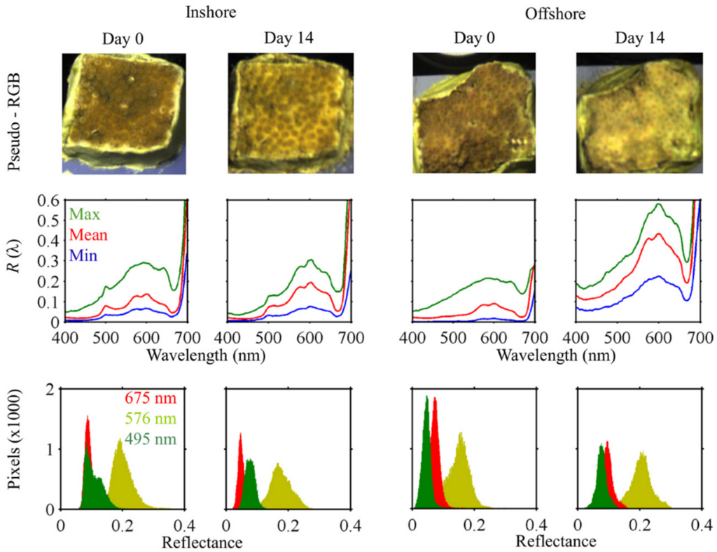
In Figure 1, the top row presents pseudo-RGB images, highlighting pigment loss in both Inshore and Offshore colonies following heat exposure, with a more pronounced effect in Offshore samples. The middle row displays mean fragment reflectance (R(λ)), showing a moderate increase for the Inshore fragment and a more significant rise for the Offshore fragment, indicative of bleaching. The bottom row illustrates shifts in pixel histograms at select wavelengths, capturing spectral changes associated with bleaching. This hyperspectral imaging approach provides a more detailed and spatially resolved assessment of reflectance variability compared to traditional fiber-optic spectroradiometry.
Why This Matters
This study demonstrates that hyperspectral imaging can serve as a powerful, non-invasive tool for monitoring coral health. Unlike conventional methods that require physical sampling and laboratory analysis, hyperspectral imaging allows for rapid, in situ assessments of large reef areas. This is particularly valuable for conservationists and marine biologists who need efficient ways to track reef degradation over time.
The Role of the SOC710-VP
The success of this research was largely due to the high spatial and spectral resolution of the SOC710-VP hyperspectral camera. Some of its key contributions included:
- High-resolution spectral mapping: Enabled researchers to capture detailed reflectance variations across different coral colonies.
- Non-invasive analysis: Allowed for real-time monitoring of coral health without disturbing fragile ecosystems.
- Broad spectral range (380–1040 nm): Covered both visible and near-infrared wavelengths, crucial for detecting bleaching and other physiological changes.
- Field-ready setup: The imager’s robust design made it ideal for use in outdoor marine environments under natural sunlight.
Future Applications
The potential applications of hyperspectral imaging extend far beyond coral monitoring. This technology could be used for:
- Early detection of reef degradation
- Mapping biodiversity in marine ecosystems
- Assessing the effectiveness of conservation efforts
- Identifying pollutants affecting oceanic habitats
With continued advancements in hyperspectral imaging, tools like the 710-VP from Surface Optics are set to play a pivotal role in the future of environmental science. As climate change accelerates, having precise, real-time data on coral health could make all the difference in protecting these vital ecosystems.
To read more about this study, you can read the original article here:
Russell BJ, Dierssen HM, LaJeunesse TC, Hoadley KD, Warner ME, Kemp DW, Bateman TG. Spectral Reflectance of Palauan Reef-Building Coral with Different Symbionts in Response to Elevated Temperature. Remote Sensing. 2016; 8(3):164. https://doi.org/10.3390/rs8030164