Oil paintings often degrade over time as many pigments and binders slowly react to damaging internal and external factors.3 When that happens, colors may fade or altogether disappear, making it harder to visualize the artist’s original intent.
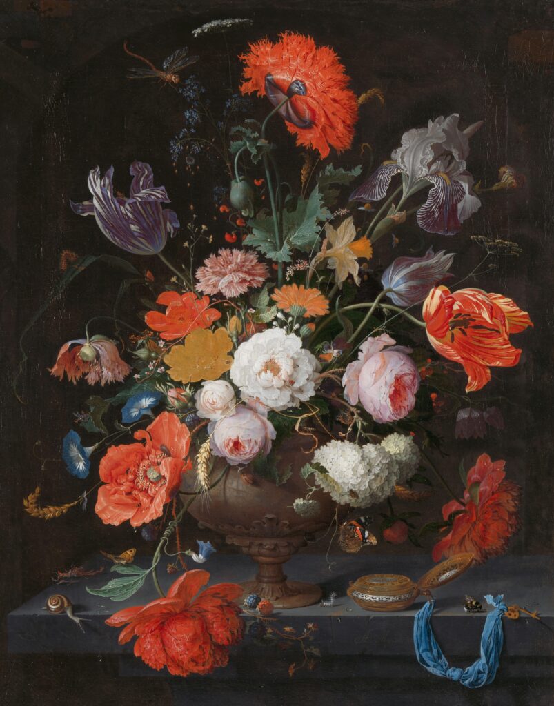
One example of this occurrence is found in Still Life with Flowers and a Watch, a 17th-century work by still-life painter Abraham Mignon on display at the Rijksmuseum in Amsterdam.2 Completed sometime between 1660 and 1679, the painting shows a yellow rose that has dulled and flattened over the years.
As a result, it lost the three-dimensional illusory effect that originally created depth through Mignon’s masterful shading techniques. The loss disrupts the visual harmony of an otherwise beautifully rendered floral arrangement.
To help unlock the hidden optics and aid in the later reconstruction of the original bloom, researchers turned to the SOC-710 Hyperspectral Camera from Surface Optics Corporation. By capturing detailed spectral and spatial data, this imaging tool played a key role in revealing what the naked eye can no longer see.
Background
Abraham Mignon’s Still Life with Flowers and a Watch showcases the technical brilliance of 17th-century Dutch still-life painting. Yet in recent decades, conservators noticed that one of its most striking elements, the yellow rose, had lost its visual impact.
Once vibrant and dimensional, the flower now appears flat, powdery, and pale. Researchers sought to understand what caused the deterioration and how the original appearance could be recovered, at least virtually.
To investigate the damage, the study combined multiple imaging and analytical tools. Elemental mapping was done with macro X-ray fluorescence (MA-XRF). Crystal structures and pigment degradation products were analyzed using macro X-ray powder diffraction (MA-XRPD). Microscale paint samples were examined under a microscope. To capture reflectance across visible and near-infrared wavelengths, the team utilized the SOC-710 hyperspectral camera1.
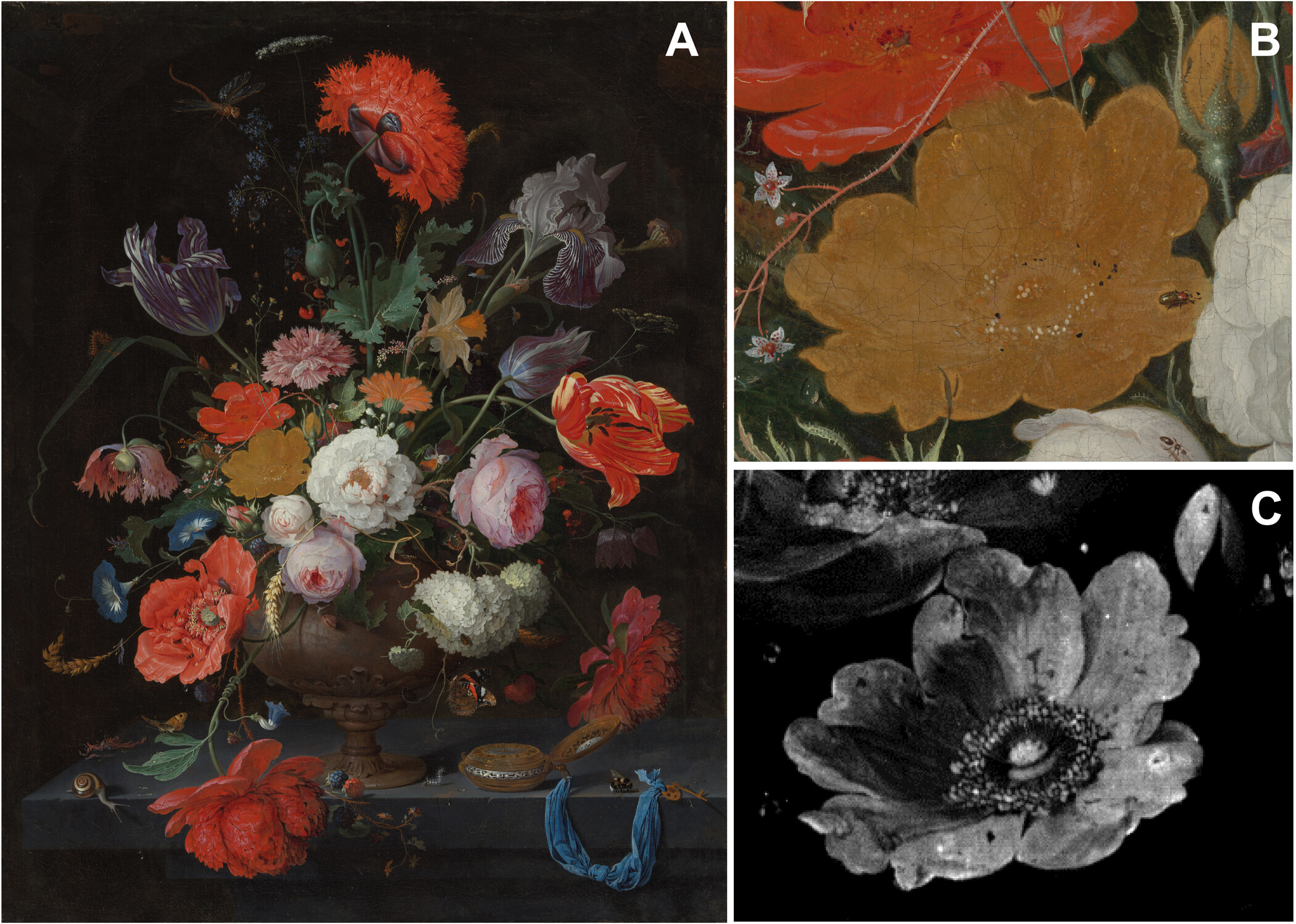
In combination, these techniques revealed that the yellow pigment orpiment had transformed into colorless arsenic compounds, including arsenolite and lead arsenates. Additionally, a transparent yellow glaze had faded due to light exposure.
The result was a loss of contrast and depth, removing Mignon’s careful light-shadow modeling. By layering insights from each method, including spectral imaging, researchers could reconstruct how the rose once looked and explain how subtle chemical changes reshape the viewer’s perception of the original.
SOC-710’s Role and Capabilities
The SOC-710 hyperspectral camera from Surface Optics Corporation played a key role in the investigation of pigment degradation in Still Life with Flowers and a Watch. Designed for laboratory and field use, the SOC-710 captures data in the visible and near-infrared (VNIR) range. The camera’s ability to record subtle changes in surface reflectance makes it especially valuable for applications in art conservation and heritage science.
One of the SOC-710’s core strengths is its internal line-scanning design, which enables precise spectral imaging without requiring external motion systems. Its compact, internally scanning design makes the SOC-710 well-suited for use on delicate or immobile subjects such as historic paintings.
In the study, the SOC-710 was used to collect reflectance data across a wide range of wavelengths. This process highlighted differences in pigment composition that were not detectable without advanced imaging. VNIR Imaging revealed that the upper paint layers had become degraded and transparent over time, exposing different underlayers containing iron-rich pigments, such as goethite.
Experimental Setup
The spatial resolution at the painting was 0.168 mm, corresponding to 172 mm field of view, and the integration time used was 100 ms. Two light sources Solux 4700K, 50-W lamps (coated to minimize UV and IR light) were used to collect the reflected signal. A step/stare collection approach was used to collect VNIR image cubes of the entire surface of the painting to allow generating a complete image cube. To do this, the camera and lights remained stationary, the painting was moved left-right and up-down on an easel, and a total of 20 cubes (1024 × 1024 pixels) were acquired to have the final VNIR reflectance data cube.1
The SOC-710 collected reflectance data in the VNIR range. These measurements revealed how pigment layers had changed chemically and optically. Researchers observed that degraded orpiment had become transparent, making iron-based pigments in the underpainting, such as goethite, more visually dominant than intended. This determination was critical in reconstructing the rose’s original three-dimensional shading and color gradients.
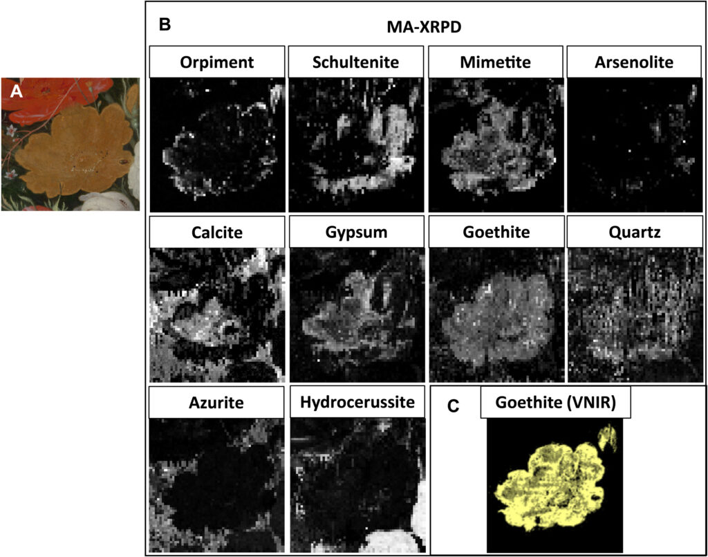
(A) Optical photograph of the analyzed area. (B) Distribution images obtained with reflection MA-XRPD, showing the intensity scaling parameter for every pixel. (C) Map obtained with the portion of the spectral endmember showing characteristic features of goethite, contained in the yellow ocher pigment.
The SOC-710’s spectral data was analyzed alongside elemental maps from macro X-ray fluorescence (MA-XRF) and crystal structure data from macro X-ray powder diffraction (MA-XRPD). The study found that MA-XRPD helped identify products that had formed within the original paint layers. Researchers could then match these compounds with visible changes in the painting, such as areas where vibrancy and contrast had been lost.
Used in combination, these techniques formed a comprehensive imaging workflow. MA-XRF revealed the distribution of elements such as arsenic and lead. MA-XRPD confirmed which mineral phases were present. The SOC-710 added a spectral dimension that linked these materials to shifts in how light interacted with the surface. Together, the imaging tools allowed researchers to digitally reconstruct the visual modeling once created by Mignon’s careful use of light, color, and pigment layering.
Results
The SOC-710 provided essential spectral and spatial data that deepened researchers’ understanding of pigment degradation in Mignon’s yellow rose.
Reflectance data from the SOC-710 revealed fine variations in pigment behavior. These patterns matched elemental maps from MA-XRF, especially those showing the distribution of arsenic and calcium. Researchers linked these findings to the original light-shadow modeling that once gave the flower its dimensional form.
The camera also distinguished spectral signatures tied to specific degradation products. In particular, it helped differentiate orpiment from arsenate compounds like mimetite and schultenite. These products formed within the original paint layers and contributed to the loss of color and contrast.
The spectral information supported digital reconstruction efforts described in the paper. By combining reflectance imaging with elemental and structural data, the researchers proposed a visual model that closely aligned with the artist’s original design. Their reconstruction showed how light and shading were intended to move across the surface of the rose.
Ultimately, the SOC-710 did more than detect material loss; it helped recover visual meaning, allowing researchers to reconnect what is physically present with what the artist once intended the viewer to see.
Broader Impacts of the SOC-710 Beyond the Conservation Lab
The SOC-710 hyperspectral camera from Surface Optics Corporation played a pivotal role in revealing how pigment degradation reshaped a key feature of a 17th-century masterpiece. Its ability to capture detailed reflectance data at the microscale helped researchers identify material changes that traditional techniques couldn’t fully explain. In doing so, it brought lost visual elements of Abraham Mignon’s formerly degraded yellow rose closer to its original vibrant form.
Beyond this single case, the SOC-710 demonstrates the broader value of non-invasive, high-precision imaging in the field of cultural heritage work. Without touching the artwork, researchers could trace chemical transformations, isolate their visual effects, and propose informed digital reconstructions.
These insights supported digital reconstructions of the painting’s original appearance. That data can also be used to generate accurate visual reproductions for use in virtual expeditions, educational programming, licensed digital editions, and similar applications across museums and academic institutions.
In addition to digital outputs, hyperspectral imaging supports physical restoration by pinpointing where degradation has occurred and revealing the material beneath altered surfaces.
For professionals such as conservators, curators, and imaging scientists, hyperspectral imaging continues to expand what is possible in the preservation and study of aging artworks. These insights can guide decisions about where and how to clean, retouch, reinforce, or stabilize vulnerable areas without compromising the integrity of the original piece.
To learn more about how hyperspectral imaging can support your research, reach out to their team at +1-858-675-7404 or contact@surfaceoptics.com for a consultation.
Sources
[1] De Keyser, N., Broers, F., Vanmeert, F., De Meyer, S., Gabrieli, F., Hermens, E., Van der Snickt, G., Janssens, K., & Keune, K. (2022). Reviving degraded colors of yellow flowers in 17th century still life paintings with macro- and microscale chemical imaging. Science Advances, 8(29), eabn6344. https://doi.org/10.1126/sciadv.abn6344.
[2] Rijksmuseum. (n.d.). Still Life with Flowers and a Watch Abraham Mignon, c. 1669–c. 1679. https://www.rijksmuseum.nl/en/collection/object/Still-Life-with-Flowers-and-a-Watch–7404dc80eb5bfc4161ed6ccf454e293f
[3] Miliani, C., Monaco, L., Fantacci, S., Romani, A., Melo, J., Angelin, E., & Janssens, K. (2018). Recent insights into the photochemistry of artists’ pigments and dyes: Toward better understanding and prevention of colour change in works of art. Angewandte Chemie International Edition. https://www.iperionhs.eu/wp-content/uploads/2018/06/Miliani_et_al-2018-Angewandte_Chemie_International_Edition.pdf
Ready to Explore Hyperspectral Imaging for Your Application?
Curious if hyperspectral imaging could work for your project? Drop us a line—we’d love to hear what you’re working on.