The need to conserve art has a long history, back to the time when people first realized that the art objects they treasured had begun to deteriorate. In the nineteenth century science and art first began to meet, with scientists of that time looking for methods to preserve valuable works of art.
Of course today many of their methods, such as varnishes, have been seen to actually contribute to, rather than prevent, the deterioration of paintings, but that early blend of art and science has continued up to today, where art conservation draws heavily on scientific methods for analysis of paintings, identification of conservation issues and the detection of additions and forgeries.
HSI APPLICATIONS
- Pigment Identification and Mapping
- Examination of Underdrawings
- Analysis of 3-D Objects
- Recognition of Organic Materials
- Forgery Detection
Traditional scientific methods include chemical analysis of paint samples to determine the molecular nature of the pigments and varnishes used. X-ray technology is also often employed to see beneath surface layers and discover the intentions and processes of the artist.
Although these methods have been employed with success for some time, many of the techniques used either give limited information, are invasive to some degree, or both.
There is still a need for techniques that will yield valuable information without damaging the work directly or exposing it to extreme light levels that may accelerate deterioration. Hyperspectral imaging has been engaged with promising results to widen the range of tools available to art conservation specialists.
Reflectance spectroscopy has been used successfully on numerous occasions to gather information for art conservation activities. Reflectance spectrophotometers have been used in combination with digital imaging to produce valuable results. These methods allow non-destructive study, but are limited by the cumbersome equipment, and the limited wavelength bands available. Additionally, their use is confined to spot-testing. Since the surfaces of painting are extremely heterogeneous, this is a severe limitation that can now be overcome with hyperspectral imaging, which is cheaper, gives a full range of spectral results and can be used to capture data from the complete artwork.[1]
Pigment Identification and Mapping
One key area in the analysis of artworks is the identification of pigments. This identification provides invaluable information on the age of a painting, improving dating and attribution. It also allows restorers to work with the correct pigments to match those used in the work being restored.
Spectroscopy has been used to identify the pigments used in an art-work for a number of years. However many pigments only begin to show differences in absorption and reflectance in the Ultra-Violet area of the spectrum, so the use of visible light has significant limitations. Multispectral imaging has been used to identify the pigments used in traditional Japanese paintings.[2] Estimates were made of the pigments used and these proved to be a good match to reference samples, but significant issues with signal noise were encountered which would be eliminated with hyperspectral imaging equipment.
Hyperspectral Imaging, sometimes called ‘reflection imaging spectroscopy’, makes the collection of data across a broad range of wavelengths practical, providing sufficient data for detailed analysis. It has been shown that even over limited wavelength ranges (650 – 1040nm) and with relatively coarse resolution (10 nm), invaluable data can be collected. The equipment is portable, so it can come to the artwork, rather than risking the transportation of fragile works to laboratories. It requires only benign light-levels and the equipment is relatively inexpensive.
However before analysis can be made, there needs to be a reliable set of reference samples of pigments, inks, resins, binders and so on, from relevant periods, that can then be used for the interpretation of analysis data-sets. In a collaborative effort between the University of Winnipeg, Winnipeg Art Gallery and the National Research Council of Canada, the Centre for Scientific and Curatorial Analysis of Painting Elements (C-SCAPE) has been established to gather and catalogue data on historic pigments and art materials.
Using these reference graphs for comparison, it is possible to identify the pigments used in a work of art without the need for invasive techniques to obtain samples for chemical analysis. This provides valuable data for authenticity and provenance investigations as well as knowledge of the pigments used by the artist.
The Kubelka-Munk Equation connects light reflectance (scattering) to absorption in effectively opaque materials, such as paint on canvas. By measuring the ratio of absorption to scattering (K/S) at each wavelength, graphs can be constructed for each pigment.[3] Although there are some limitations, Kubelka-Munk analysis can produce robust results even for pigments of varying concentrations, particle size, and when found in different binding media.[4]
In a study of Picasso’s Harlequin Musician, researchers from the National Gallery of Art and the U.S. Army Night Vision & Electronic Sensors Directorate compared the spectra collected with two Surface Optics hyperspectral cameras with those from a fiber optic reflectance spectrometer (FORS). Researchers found that hyperspectral data taken in the visible to shortwave infrared (400 – 1650nm) produces FORS quality spectra for the complete surface of the painting and could be used to separate, identify and map pigments.
Access the full article “Visible and Infrared Spectroscopy Imaging of Paintings: Pigment Mapping and Improved Infrared Reflectography” in the SPIE Digital Library.
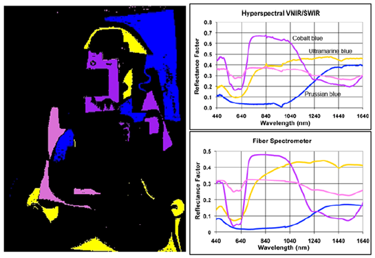
Identification and mapping of the primary blue pigments used in the Harlequin Musician obtained from the combined VNIR and SWIR image cubes and compared with Vis-SWIR FORS measurements.[5]
Examination of Under-drawings
A great deal of valuable information about the intentions of the creator of an artwork, such as changes made during the creative process, alterations and the manner in which the artist worked, can be determined from the examination of underlying drawings, sketch lines and work that was eventually painted out.
X-ray technology has been used for this purpose[6], but has limitations – for example charcoal sketch lines on dark backgrounds cannot be distinguished. Hyperspectral imaging offers the possibility of seeing more clearly through surface layers.
Light penetration is a function of wavelength. This is shown by the Beer-Lambert Law, demonstrating that penetration is inversely proportional to wavelength. This means that since ultraviolet light has a shorter wavelength it will penetrate further through paint layers to reveal what is below.[7] Wavelengths in the 1000-2000nm range give the best results.[4] Hyperspectral imaging can then capture information that reveals under-drawings, foundation materials and other information of great value to art conservationists.While single-wavelength data can be of some value, hyperspectral imaging permits a dramatic increase in information since data from across the full waveband can be simultaneously displayed. Using image visualization software to process the data cube, the reflectance at each point and wavelength can be expressed in its principle components (PCs). These can be converted into gray-scale images. By removing the main PC, lesser values are revealed, which show, for example, sketch lines, and even enable discrimination between charcoal lines and ink lines.[8]
In a study at the Winnipeg Art Gallery, Canada, a 15th century drawing, Untitled (The Holy Trinity) by Viet Hirschvogel the Elder, was analyzed using hyperspectral imaging equipment, to reveal not only the lead-pencil outline used for the drawing sketch, but the several different inks used to create the base for the actual drawing.[9] Pablo Picasso’s The Tragedy is known to have multiple compositions beneath the finished painting, identified with broadband infrared reflectography and X-ray imaging.[10] The National Gallery of Art and U.S. Army Night Vision & Electronic Sensors Directorate researchers took hyperspectral images using an 85-band Surface Optics SWIR imager and found the spectral bands at which optimal visualization of under drawings occurred.
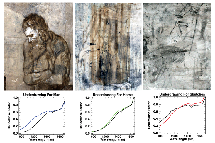
False color infrared reflectograms showing three detailed sections from The Tragedy. Each image shows a selection of the spectral bands that best reveal the various underdrawings and caricatures on the panel. Left: Drawing for the man 1000, 1150, 1200 nm; Middle: Horse 1300, 1350, 1400 nm; Right: Sketches 1600, 1625, 1660 nm.[5]
Delaney et al. (2009) explained, “Prior broad-band infrared images revealed many of these features, however, they were difficult to discern because of the low signal to noise ratio of the sensing method and the jumble of images present. Therefore, the combination of reflectance spectra and narrow band false color composite images improve the ability to emphasize features of interest when compared to the earlier methodology.”
Analysis of 3-D Objects
The versatility of hyperspectral imaging means that three-dimensional objects can also be analyzed. In a study of Michelangelo’s David, spectral ‘signatures’ from various parts of the work were taken.[4] These signatures were created by measuring changes at different wavelengths in the reflectance of a nitrogen laser shone on the sculpture. These could then be compared to the results from a sample of the original Carrara marble used to create the work. Some areas showed variations from the Carrara signature, indicating where repairs had been made.
Recognition of Organic Materials
Renaissance artists used a variety of materials to bind their pigments and often different materials were used with different pigments in the same painting. In a study of Cosimo Tura’s The Annunciation with Saint Francis and Saint Louis of Toulouse (circa 1475), it was possible to identify which binders had been used. Reference samples of the most common materials, animal skin glue and egg yolk, could be matched using near infrared hyperspectral imaging to specific areas of the painting and to specific pigments.[11] Researchers used a Surface Optics hyperspectral imager operating from 950 to 2500 nm with a spectral resolution of 4.4 nm.
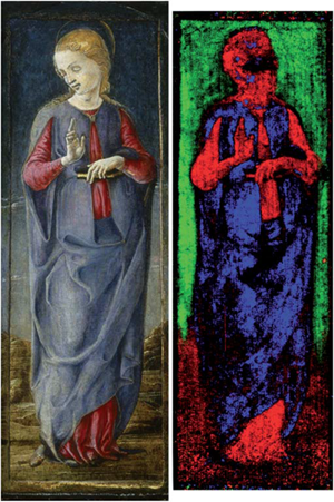
The visible image of the Virgin panel next to the false-color mapping of pigment binders. The red corresponds with egg yolk binder, the blue is consistent with a glue binder, and the green maps the areas of Azurite in a glue binder.[11]
For consultation about a particular application and information on leasing or purchasing a hyperspectral camera, complete our contact form.
1. Kubik, Maria. Hyperspectral Imaging: A New Technique for the Non-Invasive Study of Artworks. Dudley Creach and David Bradley. Physical Techniques in the Study of Art, Archaeology and Cultural Heritage, Volume 2. s.l. : Elsevier, 2007.
2. Pigment identification by analytical imaging using multispectral images. Toque, J.A., Sakatoku, Y. and Ide-Ektessabi, A. 2009. 16th IEEE International Conference on Image Processing.
3. Johnston-Feller, Ruth. Color Science in the Examination of Museum Objects: non-destructive procedures. Getty Publications, 2001.
4. Paparo, Omer. Lecture 11 – Hyperspectral Imaging applications in art and archaeology (PPPres). Haifa University. [Online] 2013. http://cs.haifa.ac.il/hagit/courses/seminars/Hyperspectral/Presentations/.
5. Visible and Infrared Spectroscopy Imaging of Paintings: Pigment Mapping and Improved Infrared Reflectography. Delaney, John K. et al. 2009. Proceeding of SPIE. Vol. 7391.
6. Kirsh, Andrea and Levenson, Rustin S. Seeing Through Paintings: Physical Examination in Art Historical Studies. s.l. : Yale University Press, 2000.
7. Douma, M. Visible & Beyond. Pigments Through the Ages. [Online] 2008. http://www.webexhibits.org/pigments/intro/visible.html.
8. Mansfield, James R. Near infrared spectroscopic reflectance imaging: a new tool in art conservation. Journal of Cultural Heritage. 2003, Vol. 4, pp. 127-136.
9. Attas, Michael, et al., et al. Spectroscopic Imaging in Art Conservation: A New Tool for Materials Investigation. Leonardo: Journal of the International Society for the Arts, Sciences and Technology. 2003, Vol. 36, 4, 304-307.
10. Hoenigswald, A., [Picasso The Early Years, 1892-1906], The National Gallery of Art, Washington D.C., 299-309 (1997).
11. A., Dooley. Kathryn, et al. Mapping of egg yolk and animal skin glue paint binders in Early Renaissance paintings using near infrared reflectance imaging spectroscopy. Analyst. 2013, Vol. 138 , pp. 4838-4848